Home

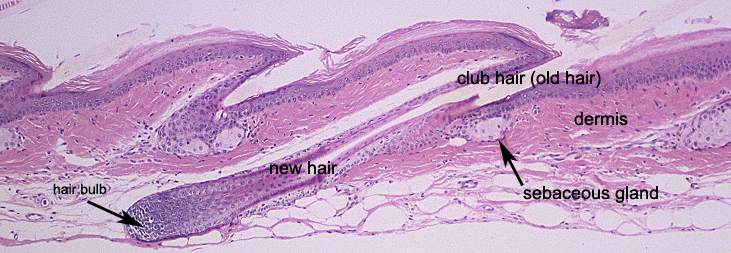
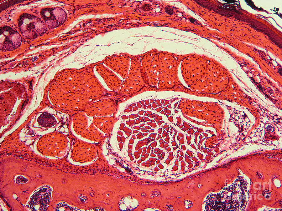
Conductividad Consumir relajarse mouse tail histology Prueba de Derbeville Dinkarville ganar

The Interfollicular Epidermis of Adult Mouse Tail Comprises Two Distinct  Cell Lineages that Are Differentially Regulated by Wnt, Edaradd, and Lrig1  - ScienceDirect
The Interfollicular Epidermis of Adult Mouse Tail Comprises Two Distinct Cell Lineages that Are Differentially Regulated by Wnt, Edaradd, and Lrig1 - ScienceDirect

Skinbase - Mutant Mouse Skin Database

Histological structure of the mouse tail skin treated with saline... |  Download Scientific Diagram
Histological structure of the mouse tail skin treated with saline... | Download Scientific Diagram

a) A stained cross-section image of a mouse tail. (b) Cross-sectional... |  Download Scientific Diagram
a) A stained cross-section image of a mouse tail. (b) Cross-sectional... | Download Scientific Diagram

Abnormal differentiation of epidermis in transgenic mice constitutively  expressing cyclooxygenase-2 in skin | PNAS
Abnormal differentiation of epidermis in transgenic mice constitutively expressing cyclooxygenase-2 in skin | PNAS
CD4+ Cells Regulate Fibrosis and Lymphangiogenesis in Response to Lymphatic  Fluid Stasis | PLOS ONE
CD4+ Cells Regulate Fibrosis and Lymphangiogenesis in Response to Lymphatic Fluid Stasis | PLOS ONE

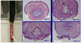
Performance of wounding experiments to test investigational agents in mouse  full-thickness tail wounds | Dermatology
Performance of wounding experiments to test investigational agents in mouse full-thickness tail wounds | Dermatology

Histology of the orthokeratotic mouse tail skin action: parakeratotic... |  Download Scientific Diagram
Histology of the orthokeratotic mouse tail skin action: parakeratotic... | Download Scientific Diagram

Histology of tail subcutis of rat and cotton rat. (A,A′) Cross-section... |  Download Scientific Diagram
Histology of tail subcutis of rat and cotton rat. (A,A′) Cross-section... | Download Scientific Diagram

Histology of the: (a) parakeratotic skin section of mouse tail and (b)... |  Download Scientific Diagram
Histology of the: (a) parakeratotic skin section of mouse tail and (b)... | Download Scientific Diagram

Blood vessels of the rat tail: a histological re-examination with respect  to blood vessel puncture methods
Blood vessels of the rat tail: a histological re-examination with respect to blood vessel puncture methods

The mechanical properties of tail tendon fascicles from lubricin knockout,  wild type and heterozygous mice - ScienceDirect
The mechanical properties of tail tendon fascicles from lubricin knockout, wild type and heterozygous mice - ScienceDirect

Mouse tail, c.s. - Laboratory equipment

Gallery : School of Biomedical Sciences

Tail of mouse (showing many tissue types) t.s. slide microscope slide

Histology of tail epidermis and dermis of rat and cotton rat. (A,A′)... |  Download Scientific Diagram
Histology of tail epidermis and dermis of rat and cotton rat. (A,A′)... | Download Scientific Diagram

Skinbase - Mutant Mouse Skin Database

Reproducibility of histopathological findings in experimental pathology of  the mouse: a sorry tail | Lab Animal
Reproducibility of histopathological findings in experimental pathology of the mouse: a sorry tail | Lab Animal

Histology of tail skin from a control mouse ( A and D ), a heterozygous...  | Download Scientific Diagram
Histology of tail skin from a control mouse ( A and D ), a heterozygous... | Download Scientific Diagram

Osteoderms in a mammal the spiny mouse Acomys and the independent evolution  of dermal armor - ScienceDirect
Osteoderms in a mammal the spiny mouse Acomys and the independent evolution of dermal armor - ScienceDirect

Histology of the orthokeratotic mouse tail skin action: parakeratotic... |  Download Scientific Diagram
Histology of the orthokeratotic mouse tail skin action: parakeratotic... | Download Scientific Diagram
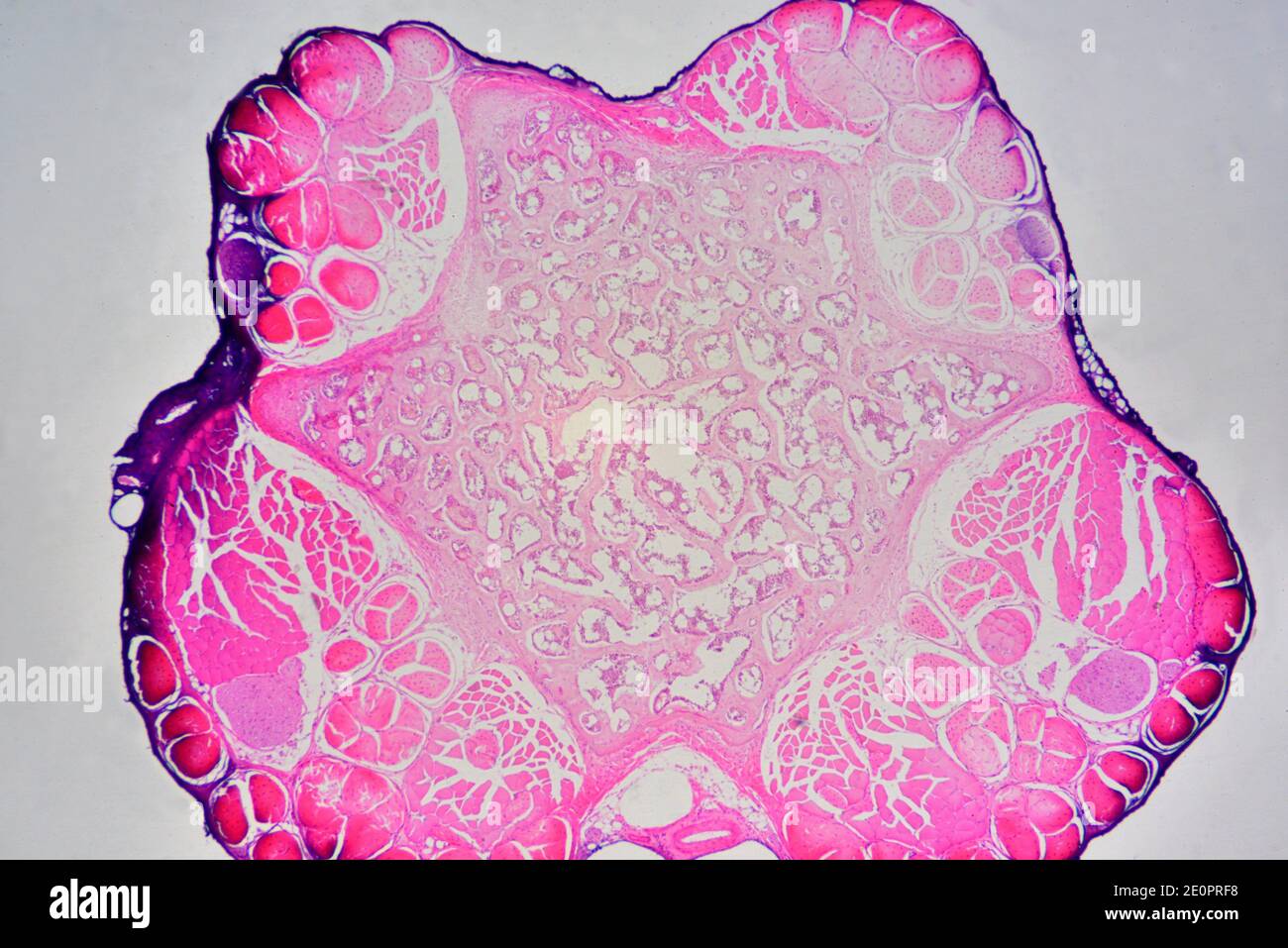
Mouse Tail Cartilage Lm Photograph by Garry DeLong - Fine Art America

Mouse tail cross section showing from outside to inside: epidermis,  cartilage, tendons, muscles, connective tissue and veins. Photomicrograph  X30 at Stock Photo - Alamy
Mouse tail cross section showing from outside to inside: epidermis, cartilage, tendons, muscles, connective tissue and veins. Photomicrograph X30 at Stock Photo - Alamy

Mouse tail internal structure, light micrograph - Stock Image - C049/8806 -  Science Photo Library
Mouse tail internal structure, light micrograph - Stock Image - C049/8806 - Science Photo Library

Reproducibility of histopathological findings in experimental pathology of  the mouse: a sorry tail | Lab Animal
Reproducibility of histopathological findings in experimental pathology of the mouse: a sorry tail | Lab Animal