Home

Premisa puntada Hervir cytoid bodies Escudriñar seta algodón

Cytoid bodies in cutaneous direct immunofluorescence examination - Wu -  2007 - Journal of Cutaneous Pathology - Wiley Online Library
Cytoid bodies in cutaneous direct immunofluorescence examination - Wu - 2007 - Journal of Cutaneous Pathology - Wiley Online Library

damsdelhi - All of the following are seen in tuberous sclerosis except? A.  Civatte bodies B. Koenen tumours C. Ash leaf macules D. Shagreen patch |  Facebook
damsdelhi - All of the following are seen in tuberous sclerosis except? A. Civatte bodies B. Koenen tumours C. Ash leaf macules D. Shagreen patch | Facebook

Light and electron microscopic studies on colloid and other cytoid bodies*

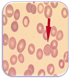
Bodies C-E - Pathological and non-pathological bodies

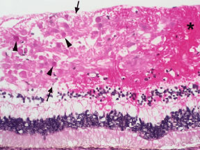
Moran CORE | Retina & RPE Histopathology

Figure 6 from Systemic lupus erythematosus and syndromes possibly related.  | Semantic Scholar
Figure 6 from Systemic lupus erythematosus and syndromes possibly related. | Semantic Scholar

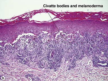
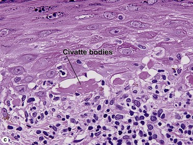
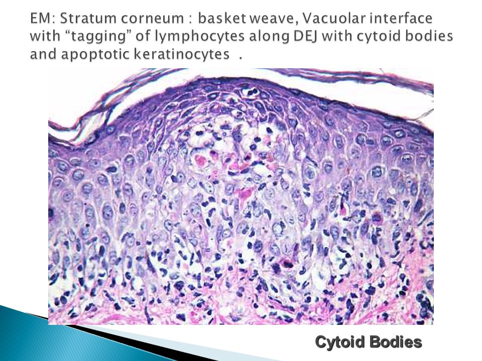
Interface dermatitis | Plastic Surgery Key

LP. DIF: Note deposition of IgM within scattered cytoid bodies in the... |  Download Scientific Diagram
LP. DIF: Note deposition of IgM within scattered cytoid bodies in the... | Download Scientific Diagram

Light and electron microscopic studies on colloid and other cytoid bodies*

Mention... - Egyptian Society of Dermatopathology - ESDP | Facebook

Figure showing skin biopsy of the lesion. Vacuolar alteration of the... |  Download Scientific Diagram
Figure showing skin biopsy of the lesion. Vacuolar alteration of the... | Download Scientific Diagram

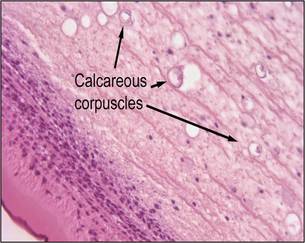
bodiesindermatology-.pptx

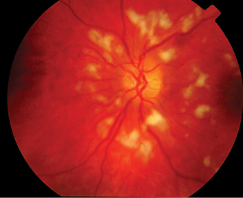
Cotton-Wool Spots May Challenge Diagnosis

Lymphohistiocytic infiltrate with occasional eosinophils in the... |  Download Scientific Diagram
Lymphohistiocytic infiltrate with occasional eosinophils in the... | Download Scientific Diagram

Differential staining of cytoid bodies and skin-limited amyloids with  monoclonal anti-keratin antibodies. | Semantic Scholar
Differential staining of cytoid bodies and skin-limited amyloids with monoclonal anti-keratin antibodies. | Semantic Scholar

Cotton-Wool Spots (Inner Retinal Ischemic Spots) | Ento Key

Bodies C-E - Pathological and non-pathological bodies

Fever of unknown origin | PPT

Interface dermatitis | Plastic Surgery Key

Fig. 22.
Fig. 22.

Lichen planus ppt | PPT

Fig. 10.
Fig. 10.

Erythema, Purpura,Urticaria, Mastocytosis, EM, SJS and TEN - ppt video  online download
Erythema, Purpura,Urticaria, Mastocytosis, EM, SJS and TEN - ppt video online download